
Core Manager
Satoru Kondo
Project Associate Professor
Mission:
The Imaging Core enables comprehensive data acquisition on brain structure and function at different spatial and temporal resolutions using our advanced optical imaging equipment.
Technology:
The Imaging Core provides the highest level of microscope equipment in the country. Observations of fixed brains, cultured cells, and even cells in the living mouse brain can be examined with various spatio-temporal resolutions ranging from submicron to whole brains.
Description / Pre-training course application / Reservation
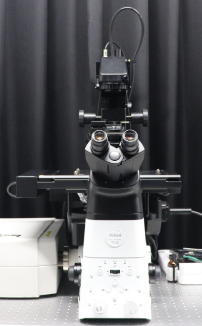
Confocal microscope Nikon A1R
It is an inverted type confocal laser microscope system. This microscope has a large FOV size of 25 and is equipped with a resonant scanner and laser unit LU-N4 (405, 488, 561 and 640 nm), and GaAsP detector. They enable to obtain images with high-speed and high-sensitivity. In addition, culture system (made by Tokai Hit) is also provided, and long-time live cell imaging is possible.
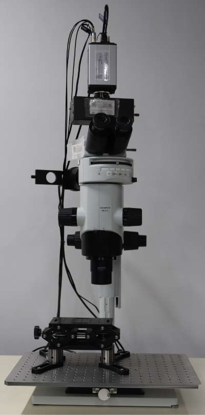
Macro zoom microscope OLYMPUS MVX-10
https://www.olympus-lifescience.com/en/microscopes/macro/mvx10/
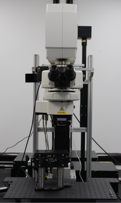
Two photon microscope Nikon A1RMP2
It is an upright type multiphoton laser microscope system. High sensitivity and high quality images can be acquired with the GaAsP NDD unit. By using piezo elements to drive the objective lens in the Z-axis direction, it is possible to take 3D images at high speed. The femtosecond pulse laser can simultaneously use two laser lines with a variable wavelength of 700-1300 nm and a fixed wavelength of 1040 nm, and is suitable for long-wavelength dye and multicolor imaging.
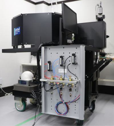
Virtual reality system PHENOSYS Jet Ball VR system
Virtual Reality (VR) systems can be used with the mouse to investigate brain function during behavioral tasks. You can examine the basic mechanisms of animal navigation, recognition, learning, or memory combined with two-photon and wide-field microscopes.
![Patch Clamp system [Patch clamp amplifier]Sutter Instruments IPA,[Manipulator]Sutter Instruments TRIO-245](../../wp-content/themes/IRCN/assets/img/archive/imaging-core/img_patch-clamp.png)
Patch Clamp system [Patch clamp amplifier]Sutter Instruments IPA,[Manipulator]Sutter Instruments TRIO-245
The patch clamp amplifier supports dual recording. In addition, the manipulator can be driven in 3 axis directions + diagonal direction. We are preparing for in vivo patch clamps, so you need to prepare by yourself for performing in vitro experiments such as slice patches.
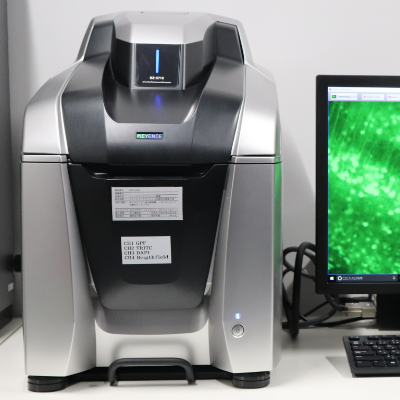
All in one microscope KEYENCE BZ-700
It is a fluorescence microscope that does not require a dark room. With the sectioning function, you can obtain clear images without fluorescent blur with easy operation. In addition, high resolution wide field of view image can be taken at high speed by image joint function.
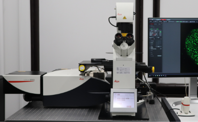
Super resolution STED microscope Leica TCS SP8 STED 3x
https://www.leica-microsystems.com/products/confocal-microscopes/p/leica-tcs-sp8-sted-one/
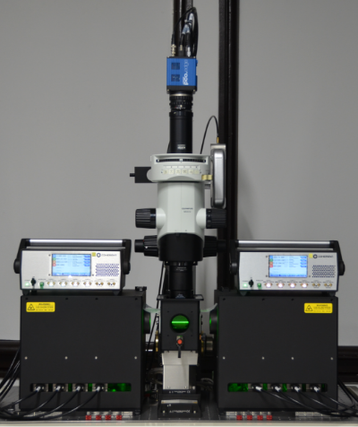
Light sheet microscope RAPID
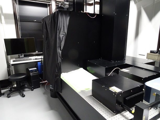
Rapid-Scope is a custom-designed light-sheet fluorescence microscope (LSFM) with mesoscopic magnitude (0.5X-2X) to explore a volumetric sample by less-invasive optical slicing. The cleared brain sample is dipped in the oil and illuminated by spatially-uniform thin light-sheet which provides a three-dimensionally isotropic resolution and covers a wide area of visualization.
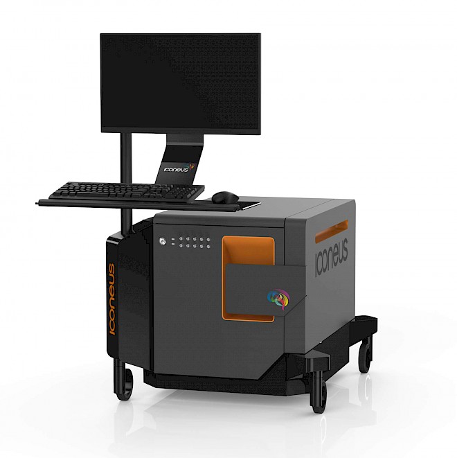
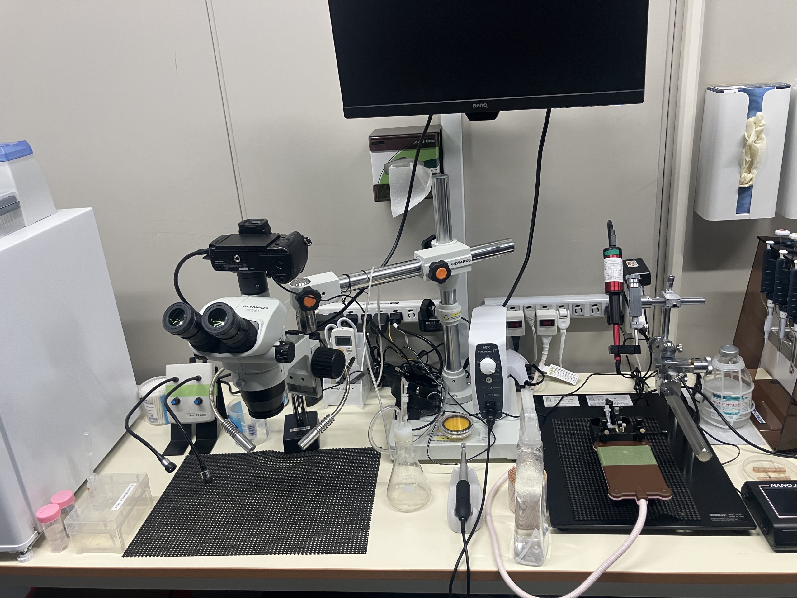
Treatment table for mice and rats
List of scheduled events
There are currently no scheduled events.
Past event list
-
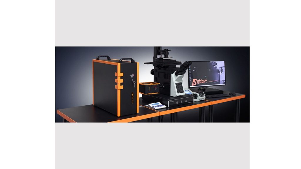 April 26th (Tue) -April 28th (Thu), 2022 Morning 10:00-12:00 Afternoon 13:00-17:00 1 hour each Super-resolution microscope "STEDYCON" demons...More
 -
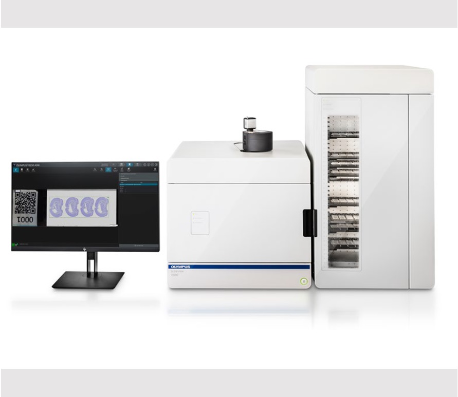 2021/11/25~2021/12/10 ①10:00-12:00 ②14:00-16:00 OLYNPUS VS200 DemonstrationMore
 -
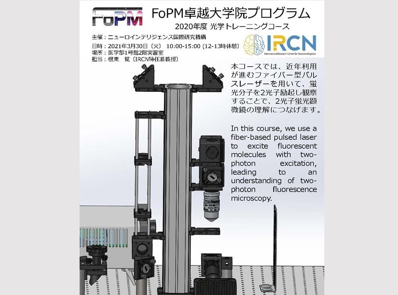 March 30, 2021 10:00-15:00 Optical Training Course 2020(FoPM Program)More
 -
 14:00 - 16:00, Monday 17. February, 2020 The 10th IRCN Imaging Core Optic technical se...More
 -
 14:00 - 16:00, Wednesday 5. February, 2020 The 9th IRCN Imaging Core Optic technical sem...More
 -
 14:00 - 16:00, Wednesday 18. December, 2019 The 8th IRCN Imaging Core Optic technical sem...More
 -
 14:00 - 16:00, Wednesday 27. November, 2019 The 7th IRCN Imaging Core Optic technical sem...More
 -
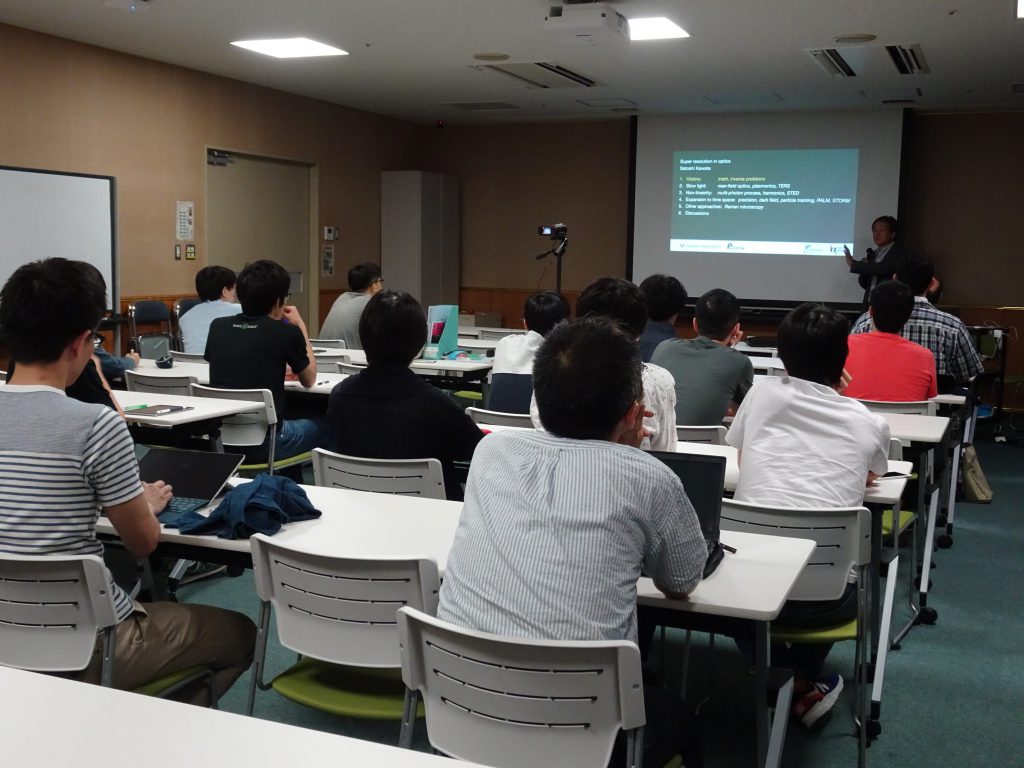 July 10, 2019 The 12th Optics Basic seminar “Super resoluti...More
 -
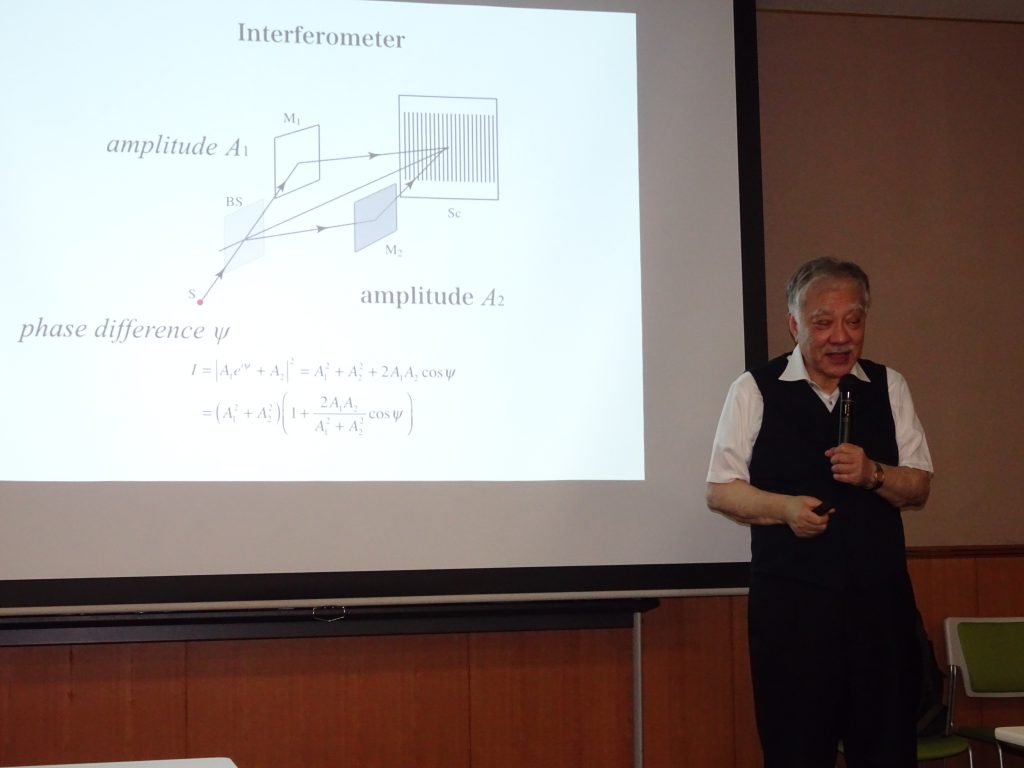 July 3, 2019 The 11th Optics Basic seminar “Nonlinear Opti...More
 -
 June 26, 2019 The 10th Optics Basic seminar “Polarization”More
 -
 June 19, 2019 The 9th Optics Basic seminar “Interference”More
 -
 June 12, 2019 The 8th Optics Basic seminar “Diffraction”More
 -
 June 5, 2019 The 7th Optics Basic seminar “Aberration2”More
 -
 May 29, 2019 The 6th Optics Basic seminar “Aberration”More
 -
 May 22, 2019 The 5th Optics Basic seminar “Optical Device”More
 -
 May 8, 2019 The 3rd Optics Basic seminar “Geometrical opt...More
 -
 May 15, 2019 The 4th Optics Basic seminar “Geometrical opt...More
 -
 April 10, 2019 The 2nd Optics Basic seminar “Light propagati...More
 -
 April 11, 12, 2018 The 2nd Optic Training Course “CameraTraining...More
 -
 April 4, 2019 The 1st Optics Basic seminar “Basic nature of...More
 -
 March 15, 2018 The 1st Optic Training Course “Spatial Proper...More
 -
 March 13, 2018 The 6th Technical Seminar “Building a Light-S...More
 -
」-min-1024x768.jpg) February 28, 2018 The 5th Technical Seminar “Optical system of ...More
February 28, 2018 The 5th Technical Seminar “Optical system of ...More -
 February 7, 2018 The 4th Technical Seminar “PMT (Photo Multi T...More
 -
 December 7, 2018 The 3rd Technical Seminar “Spatial light modu...More
 -
 November 12, 2018 The 2nd Optic Technical seminar “Basics and n...More
 -
 October 18, 2018 The 1st Optic Technical seminar “The state-o...More
Past events are here
Usage Application Form
Carry-in laboratory equipment application form
Terms of Use
Rules of the microscope room usage
Important Notice Regarding the Observation of Fixed Samples
Rules for bringing animals to Imaging Core
Microbiological monitoring items
Pledge -Ultra-sound-Imaging-system
Pledge -Ultra-Wide-Two-photon-microscope
